Radiation Survey Meters: A Comprehensive Guide
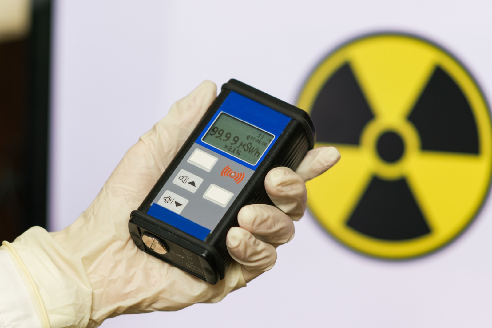
Learn about the evolution and types of radiation survey meters for ensuring safety in healthcare.
For over a century, ionizing radiation has been an integral tool in healthcare, saving lives through radiation therapy and giving providers unprecedented visual insight into the human body. It’s easy to agree that modern healthcare would not exist without radiation.
However, radiation is invisible, silent, and odorless, making it incredibly difficult to track. Accidental radiation exposure is high on the list of potential hazards in healthcare environments, especially with the introduction of nuclear medicine and advanced imaging techniques that require an injection of radioactive material. Therefore, healthcare facilities rely on specialized devices called radiation survey meters to act as their eyes and ears for potential spills and contamination. As technology advances, so do the capabilities of radiation survey meters, with modern devices offering more precise readings, greater ease of use, and enhanced safety features.
What is a Radiation Protection Survey?
A radiation protection survey is a comprehensive evaluation conducted to assess the levels of ionizing radiation in a particular area or environment. This assessment ensures that individuals working with or around radioactive materials are not exposed to harmful radiation. Radiation survey meters measure the intensity and distribution of radiation, identifying areas where radiation levels exceed safe limits.
Radiation protection surveys have two main objectives:
- 1. Ensure that all operations involving radioactive materials comply with safety standards and regulations, minimizing the risk of radiation exposure to workers and the public.
- 2. Identify the location, scale, and type of radiation hazard, enabling the implementation of appropriate control measures, such as shielding, to protect against radiation exposure.
These surveys are typically conducted by trained radiation safety technicians, health physicists, or radiation safety officers with the expertise to interpret the data collected and recommend protective measures.
Types of Radiation Survey Meters
Radiation survey meters measure and monitor levels of ionizing radiation to ensure environmental safety, personal protection, and compliance with regulatory standards. These devices vary in complexity, sensitivity, and intended application, making it essential to understand the different types available on the market. Here, we explore the primary categories of radiation survey meters, highlighting their unique features and typical uses.
Geiger-Muller (GM) Counters

GM counters are some of the most recognized radiation detection instruments. They are simple to operate but vary in effectiveness.
A Geiger counter consists of the primary radiation detection component, the Geiger-Müller tube, and a processing unit. The GM tube is a gas-filled enclosure with a central wire electrode, the anode, surrounded by an outer conductive tube, the cathode. The tube is typically filled with a noble gas (like helium, neon, or argon) mixed with a halogen to prolong the tube's life.
Principle of Operation
- 1. Ionization Process: When radiation enters the GM tube through a thin window, it ionizes the gas molecules, creating positive ions and free electrons.
- 2. Avalanche Effect: A high voltage between the central wire (anode) and the outer tube (cathode) creates an electric field. When radiation ionizes the gas, the free electrons accelerate toward the anode, and the positive ions move toward the cathode. As the electrons move toward the anode, they gain sufficient energy to ionize more gas molecules. This leads to a chain reaction or "avalanche" of ionizations triggered by a single radiation event.
- 3. Detection and Measurement: An electronic circuit connected to the GM tube detects the current generated by collecting electrons and charged ions on the electrodes. Each pulse corresponds to radiation-induced ionization within the GM tube. The processing unit counts these pulses over time, providing a measurement of the radiation intensity. The count rate (pulses per second) can be displayed directly as radiation levels using units such as counts per minute (CPM) or can be converted into dose rates (e.g., microsieverts per hour, μSv/h) using calibration factors specific to the type of radiation.
Applications and Limitations
Geiger counters are widely used for their simplicity, durability, and ability to detect various types of ionizing radiation. However, they do not provide information on the energy of the detected radiation, making them less useful for applications requiring detailed radiation spectroscopy.
- Quenching: The GM tube must be "quenched" or reset after each detection event, neutralizing the ions and electrons to stop the current and make the tube ready to detect the next event. The halogen gases (such as fluorine) in the tube help with this process by capturing electrons and assisting in de-ionization. This brief period during the collection of the radiation-induced electrical signal is called “dead time” and limits the maximum count rate the device can accurately measure.
- Plateau Region: Geiger counters operate effectively within a specific voltage range, known as the "plateau region," where the count rate remains relatively constant with changes in voltage. Operating the counter within this region ensures stable and reliable measurements.
Scintillation Counters

As their namesake implies, scintillation counters work based on the principle of scintillation, where certain materials emit light (or scintillate) when excited by ionizing radiation.
A scintillation counter consists of two main components: a scintillator and a photodetector. The scintillator is a material that emits photons (light) in response to ionizing radiation. The photodetector converts the light into an electrical signal that can be measured and quantified.
Principle of Operation
- 1. Scintillation Process: When ionizing radiation enters the scintillator material, it interacts with the atoms, exciting and elevating electrons to higher energy states. As these excited electrons return to their ground state, they release energy in the form of light photons. The amount and wavelength of the light photons depend on the type of scintillator material and the energy of the incoming radiation.
- 2. Detection of Light by Photodetector: The light photons emitted by the scintillator are then directed toward a photodetector, typically a photomultiplier tube (PMT). The PMT has a photosensitive surface (photocathode) that converts the photons into electrons. These electrons are multiplied through a series of dynodes, leading to a measurable electrical pulse.
- 3. Signal Processing and Measurement: Each electrical pulse corresponds to a detection event of ionizing radiation. The pulses are counted and displayed as a count rate (e.g., counts per minute, CPM) or, with proper calibration, as a specific activity of the radioactive source (e.g., becquerels, curies) or dose rate (e.g., microsieverts per hour, μSv/h).
Types of Scintillator Materials
Scintillator materials can be inorganic or organic, each with its unique properties and applications:
Inorganic Scintillators: These are usually crystals, such as sodium iodide (NaI) or NaI doped with thallium (NaI(Tl)), which are highly efficient at converting gamma radiation into visible light. They are widely used in gamma spectroscopy due to their high density and atomic number, which enhance their interaction with gamma rays.
Organic Scintillators: These can be either solid or liquid and are primarily used for detecting charged particles like alpha and beta radiation. Organic scintillators are less dense than inorganic ones, making them less effective for gamma-ray detection but excellent for applications requiring fast response times and detecting low-energy particles.
Applications and Limitations
Scintillation counters are more sensitive and have better energy quantification than Geiger counters. By analyzing the amplitude of the pulses, it’s possible to perform spectroscopic analysis, identifying specific isotopes and quantifying their activity. This makes scintillation counters valuable in various applications, from nuclear medicine and research to environmental monitoring and security. However, alongside these advantages, there are some disadvantages to consider:
- Complexity and Cost: Scintillation counters are generally more complex and expensive than GM counters. The photomultiplier tubes and sophisticated electronic circuitry for signal processing contribute to the higher cost and complexity of operation and maintenance.
- Light Yield Variation: The efficiency of light production (light yield) and the response of scintillation counters can vary significantly with the type and energy of the radiation detected. This variation requires careful calibration and can complicate the interpretation of results, especially when detecting low-energy or weakly interacting radiation.
- Quenching: Organic scintillators, in particular, can suffer from quenching—a decrease in light output with increasing ionization density. Quenching can affect the accuracy of measurements, especially at high radiation intensities.
- Sensitivity to Environmental Conditions: Scintillation counters can be sensitive to environmental conditions, such as temperature and humidity, affecting the scintillator's light yield and the photomultiplier tube's performance.
Pressurized Ionization Chambers

Pressurized ionization chambers measure the change in air's electric charge caused by radiation. They are known for their accuracy over a wide range of radiation levels and their capability to measure high-energy gamma radiation. Applications include hospital radiology, nuclear medicine, radiation therapy departments, nuclear power plants, and radiographic testing environments.
Principle of Operation
- 1. Ionization Process: Like GM counters, the pressurized ionization chamber consists of a gas-filled chamber containing two electrodes: an anode and a cathode. The chamber is often filled with air, but other gases can be used for specific applications and are pressurized up to 8 atm to improve its sensitivity to radiation. When radiation enters the chamber, it interacts with the gas molecules, ionizing them. This process produces ions and free electrons within the gas.
- 2. Collection of Charge: A voltage between the two electrodes creates an electric field within the chamber. This field causes the positive ions to move toward the cathode and the electrons toward the anode. These charge carriers create an electrical current that can be measured.
- 3. Current Measurement: The current generated is directly proportional to the amount of ionizing radiation passing through the chamber. The device quantifies the radiation exposure rate by amplifying and measuring this current.
Applications and Limitations
While pressurized ionization chambers are valuable tools for measuring radiation dose and dose rate, they have limitations:
- Energy Dependence: The response of an ionization chamber can vary with the energy of the incident radiation, requiring calibration for different types of radiation and energy levels.
- Limited Range: Ionization chambers are most effective within a specific range of radiation intensities. The chamber can become saturated at very high intensities, while at very low intensities, the signal may be too weak to measure accurately.
- Mobility and Size: Some ionization chambers, especially those designed for high accuracy and sensitivity, can be bulky and unsuitable for portable use. This can limit their application in field measurements.
- Environmental Influences: Environmental conditions, such as temperature, can affect the accuracy of ionization chambers. To mitigate these effects, special design considerations or corrections are required.
Semiconductor Detectors
Semiconductor detectors leverage the electrical properties of specific elements to detect and measure ionizing radiation. Unlike gas-filled detectors like ionization chambers and Geiger-Müller tubes, semiconductor detectors operate by utilizing the ionization effects within solid-state materials. They are highly sensitive and can provide detailed energy information about the detected radiation.
Principle of Operation
- 1. Ionization in Semiconductor Material:
Semiconductor detectors are made from materials like silicon (Si), germanium (Ge), or cadmium telluride (CdTe) doped with impurities. When ionizing radiation enters the semiconductor material, it interacts with the electrons in the valence band, providing enough energy for them to jump to the conduction band, thus creating electron-hole pairs (i.e., an electron in the conduction band and a hole in the valence band where it once was).
- 2. Charge Collection:
A voltage across the semiconductor material creates an electric field and causes the free electrons to move toward the positive electrode and the holes toward the negative electrode. This movement of charge carriers generates an electrical signal proportional to the energy of the incident radiation.
- 3. Signal Processing:
The electrical signals are then amplified and processed to measure the intensity and energy spectrum of the radiation. Quantifying the energy of the incoming radiation allows for detailed spectroscopic analysis, identifying specific radionuclides based on their characteristic energy peaks.
Applications and Limitations
Semiconductor detectors are used in high-precision applications requiring detailed energy resolution and sensitivity. Radiation spectroscopy can be performed using a semiconductor detector to identify and quantify specific radionuclides based on their energy spectra. These techniques are critical in nuclear physics, environmental science, and homeland security.
Semiconductor detectors are also integral to modern medical imaging. They are used in gamma cameras and positron emission tomography (PET), producing high-resolution images of the distribution of radioactive tracers within the body. Environmental monitors and the nuclear energy industry also frequently use semiconductor-based detection technologies.
However, semiconductor detectors have certain limitations:
- • Temperature Sensitivity: Most semiconductor materials are sensitive to temperature variations, which can affect their performance. Detectors often require cooling with liquid nitrogen to reduce noise and improve resolution. CdTe-based detectors are a notable exception, operating at room temperature.
- • Radiation Damage: Semiconductor detectors can suffer from radiation damage over time, especially when exposed to high radiation doses. This damage degrades their performance, necessitating periodic calibration or replacement.
- • Cost: Compared to gas-filled detectors, semiconductors are generally more expensive regarding initial investment and maintenance (such as cooling systems for germanium detectors).
- • Complex Electronics: The operation of semiconductor detectors requires sophisticated electronics for signal amplification and processing, which increases the detection system's complexity and cost.
Despite these limitations, the superior resolution and sensitivity of semiconductor detector-based radiation survey meters make them essential tools for detailed radiation analysis. Their ability to provide precise spectral information about the detected radiation has revolutionized various applications, from medical diagnostics to environmental monitoring.
Cadmium Zinc Telluride (CZT) detectors, like the 3D CZT technology used by M3D, are known for their compact size, room-temperature operation, and excellent energy resolution. These characteristics make them ideal for precision healthcare safety and medical imaging applications.
Visualize Radiation With An Imaging Survey Meter

Imaging survey meters combine radiation detection materials with advanced imaging capabilities. These devices measure radiation levels and provide visual representations of the radiation field, enabling precise localization and identification of radioactive sources. This feature is particularly beneficial in complex environments like hospitals and healthcare facilities, where assessing the presence and distribution of small amounts of radiation across high-touch surface areas is crucial.
Unlike traditional survey meters that provide numeric data on radiation levels, imaging survey meters incorporate visual imaging techniques to create a spatial map of radiation intensity. Advanced algorithms convert raw data from the detector into meaningful images. Color coding is frequently used to represent different intensities, making it easier to identify areas of concern.
Many imaging survey meters are equipped with digital displays and interfaces, allowing for real-time visualization of radiation data. Some models also feature connectivity options, such as Wi-Fi or Bluetooth, enabling data transfer to computers or mobile devices for further analysis. This digital integration enhances the device's utility, facilitating detailed assessments and long-term monitoring.
The ability to visually map radiation sources revolutionizes safety inspections and environmental assessments. For instance, in healthcare facilities, imaging survey meters can quickly identify unintended radiation leaks or pinpoint the location of radioactive materials, ensuring patient and worker safety.
Selecting the Right Meter for Your Needs
Choosing the appropriate radiation survey meter balances several factors, including the specific type of radiation to be measured, the required sensitivity and resolution, the intended application, and budgetary constraints. Here are helpful helpful tips to get you started:
- 1. Assess Your Requirements: What is your facility's primary objective of radiation measurement? Determine whether you need to detect a specific type of radiation (alpha, beta, gamma, X-ray) or a range of radiation types. Consider the expected radiation levels and the importance of energy resolution in identifying isotopes.
- 2. Know Your Options: For healthcare facilities, especially those involved in nuclear medicine or radiation therapy, meters with high sensitivity and precision, such as those based on 3D CZT technology, may be preferable.
- 3. Develop a Budget: While advanced imaging survey meters offer significant benefits in sensitivity and visualization, they may also have a higher price tag. Weigh the cost against the potential benefits in operational efficiency, safety improvements, and compliance with regulatory standards.
- 4. Prioritize Ease of Use and Training Requirements: The level of training required for effective operation should align with the skills and expertise of the intended users. A good user interface will encourage better integration into existing workflows.
Ensure Safety and Compliance with M3D's Advanced Imaging Technology
Selecting the proper radiation survey meter is a critical decision that impacts the effectiveness of radiation safety and monitoring protocols. We understand that each healthcare facility has unique challenges and requirements regarding radiation safety.
That's why we invite you to contact us for a personalized consultation.
Our knowledgeable team is ready to discuss your needs and help you determine the best M3D imaging solution for your facility. With our expertise and advanced technology, you can enhance your radiation safety protocols, ensuring your staff's and patients' well-being while maintaining compliance with the highest industry standards.
More from the blog
3D CZT: The Future of Radiation Detection
Modern radiation detection plays a crucial role in healthcare, safeguarding patients and professionals by monitoring and managing exposure levels. arrived in fits and spurts – reactionary inventions to major events li

Revolutionizing Radiation Detection Safety with M3D
Walk into any major medical facility in the world and chances are that you’ll quickly come across a Radiology or Nuclear Medicine department full of modern medical imagers. From mammograms to CT and PET scanners, it’s no

M3D Receives NSF SBIR Phase I Grant to Advance Gamma Camera Design
The team at M3D is thrilled to announce a $275,000 SBIR Phase I grant from the National Science Foundation (NSF) to support our innovative research on gamma camera technology. This award, coupled with $25,000 in matching